General Overview

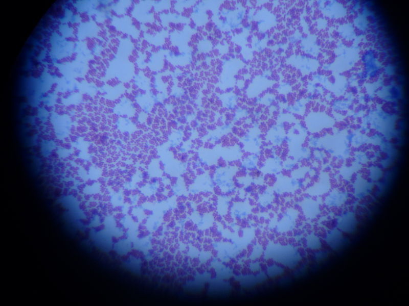
S. agalactiae is also called as group B streptococci in Lancefield grouping.

S. agalactiae found in normal flora of female genital tract, urethral mucous membranes of males and in the gastrointestinal tract (mainly in colon) as asymptomatic carriers.

However, it can cause severe invasive infections especially in newborns, elderly, and immunocompromised people. It mainly causes sepsis and meningitis in neonates which should be consider and take immediate actions.

s.agalactiae

Previously S. agalactiae recognized as a cause of bovine mastitis. However, since 1960 it has become the main cause of neonatal sepsis in industrialized countries and it is an important cause of morbidity among peripartum women and other adults with chronic medical conditions.

S agalactiae is one of most frequent isolate from blood cultures. This organism can be transmitted among adults, sexually and from an infected mother to her new born baby. High vaginal swabs, cervical swabs, umbilical swab, eye swabs, blood, or spinal fluid can be used for culture.

Pathogenesis

capsule demonstration

S. agalactiae produces many virulence factors. They are capsule polysaccharide, haemolysins, C5a peptidase, hyaluronidase and various surface proteins. There are ten different types of capsular polysaccharides.

CAMP factor is one of known haemolysin produced by S. agalactiae. Risk for neonatal diseases are premature rupture of membranes, prolonged labour, premature delivery, low birth-weight, intrapartum fever.

Infections in adults occur due to abortions, chorioamnionitis, post-partum sepsis (endometritis) and other infections.

agalactiae is a frequent cause of infection in patients with diabetes mellitus, liver cirrhosis, renal failure, stroke and cancer. Disease may manifest as pneumonia, sepsis, cellulitis arthritis, and urinary tract infections.

agalactiae is a gram positive coccii in gram stained slides and usually seen as short chains. The organisms are non-motile and most organisms are capsulated. Moreover, organism is catalase positive.

Blood agar

Most strains of S. agalactiae produce grey colour, mucoid colonies.

The diameter is about 2 mm.

Colonies show narrow zone of beta haemolysis.

About 5% of strains are non haemolytic.

Streptococcus agalactiae

Chocolate agar

The colonies are 2 mm in diameter and gray white in colour.

MacConkey agar

Most strains grow on MacConkey agar.

Neomycin blood agar

A selective medium for isolating S. agalactiae from urogenital specimens.

S. agalactiae is bacitracin resistant (0.04 IU). CAMP test is useful to confirm the organism when streptococcal grouping kit is not available.

This organism is CAMP positive.

The CAMP factor can lyse sheep or bovine red blood cells.

When it is pretreated with S. aureus the β-toxins .

Hippurate hydrolysis test also useful when streptococcal grouping kit is not available.

They hydrolyze hippurate, an important diagnostic criterion.

Positive CAMP test.

Latex agglutination tests can demonstrate the presence of group B antigen in these samples. Group B antiserum in streptococcal grouping kit gives agglutinations with positive samples.

Treatment

antibiotic treatment

Drug of choice for the treatment of S agalactiae is penicillin G. Ampicillin also useful. Aminoglycoside can be added In life-threatening occations.

Pregnant carriers under risk of premature rupture of membranes or prolonged labour should be treated with ampicillin during labour. 

Other important features

Lorem ipsum dolor sit amet, consectetur adipiscing elit. Ut elit tellus, luctus nec ullamcorper mattis, pulvinar dapibus leo.

No responses yet

Leave a Reply