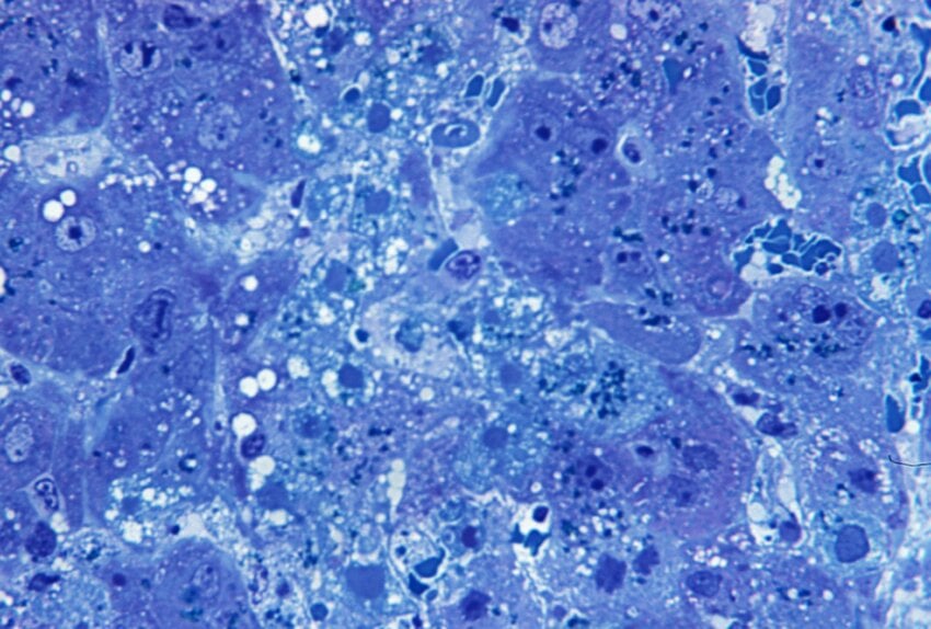
Azure A is a small planar cationic dye.

It typically stains tissues in blue colour. Methylene blue, toluidine blue are also similar dyes to Azure A.

Under metachromasia conditions, these dyes stain tissue components in purple-red colour.

These dyes are useful for the identification of charged mucins and proteoglycans.

This is one of the old histochemical staining techniques for carbohydrates.

Principle

The anionic groups of carbohydrates bind with the cationic dye molecules.

Structure of carbohydrates act as a template to induce the formation of the polymeric dye structure.

To form the polymeric structure, dye molecules bind one another via hydrogen bonds or van der Waals’ forces.

For the metachromatic phenomena integration of water molecules between adjacent dye molecules is essential.

Method

  • For the preparation of Azure A solution, dissolve 0.01 g of Azure A in 100 ml of 30% Ethanol.
  • Then dewax the sections and bring the sections down to the water through graded alcohol solutions.
  • Then treat with Azure A solution by covering the section with Azure A for 10 minutes.
  • Next rinse with distilled water.
  • After that Dehydrate the sections using graded alcohol solutions.
  • Clear with xylene.
  • Finally mount with miscible mounting medium and observe under microscope.

Results

Acid mucins and proteoglycans stains in purple to red colour. Background tissues can be seen in blue colour.

metachromasia-generating techniques such as Azure A technique have largely been replaced by techniques such as Alcian Blue technique.

Furthermore, for the demonstration of weakly metachromatic acid carbohydrate containing molecules 0.1% Azure A solution can be used.

No responses yet

Leave a Reply