Anemia is present when the red blood cells or haemoglobin level in the blood is below the lower limit for the age and the sex of the individual.

In order to diagnose anemia a complete blood count is done. Under that a blood picture of the patient is examined.

The blood picture shows the abnormalities of blood cells of the patient.

Most of the anemia types show special characteristics in the blood picture that help the diagnostic procedure.

Iron Deficiency Anemia

Iron deficiency anemia is belongs to microcytic, hypochromic anemias.

Then the blood picture of a patient who has iron deficiency anemia shows microcytic red blood cells many of which have thin rim of pink heamoglobin (hypochromia).

Red blood cells which are unequal in size (anisocytosis), poikilocytes target cells, tear drop cells and pencil shaped cells are also present occasionally.

Sideroblastic anemia is also belongs to the microcytic, hypochromic anemias.

There are some special features can be seen in the blood picture. The most evident feature is ring sideroblasts.

Immature red cells will contain iron deposited in a ring around their nuclei. Red blood cell dimorphism and basophilic stippling, which is caused by the degraded ribosomes which indicates ineffective erythropoiesis can be seen.

Iron depositions inside mitochondria (pappenheimer bodies) are also in the peripheral blood smear.

Vitamin Deficiency Anemia

This type of anemia is also called megaloblastic anemia. The deficiency of  vitamin B12 or folic acid is the main cause for vitamin deficiency anemia.

The peripheral blood picture of a patient who has vitamin deficiency anemia shows oval enlarged red blood cells (macrocytes) and hypersegmentated neutrophils.

Neutropenia and thrombocytopenia can be seen due to due to ineffective neutrophil and platelet production.

Haemolytic Anemias

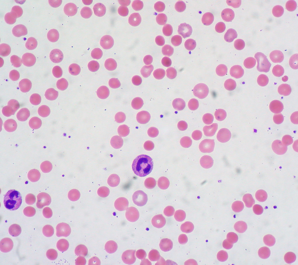
Hemolytic anemia appears when red blood cells are destroyed faster than they’re going to be made.

The destruction of red blood cells is being called hemolysis. Hemolytic anemia divides into two main categories called hereditary and acquired.

There are many types of haemolytic anemias such as hereditary spherocytosis, hereditary elliptocytosis, sickel cell anemia, thalassaemia, G6PD deficiency, pyruvate kinase deficiency.

Hereditary Spherocytosis

People with hereditary spherocytosis disorder have red blood cells that are round (spherocytes) instead of the typical biconcave shape.

These cells break down easier under stress than normal red blood cells (osmotic fragility) leading to haemolytic anemia.

Hereditary Elliptocytosis

People with hereditary elliptocytosis disorder have elliptical-shaped erythrocytes (elliptocytes) that different from the normal biconcave shape.

This condition reduce the elastic ability of the red blood cell and shortens the survival causing haemolytic anemia.

Sickel Cell Anemia

The blood picture of patients who has this condition shows multiple sickle cells.

There are also findings consistent with functional asplenia, including a nucleated red blood cell, a red blood cells containing a Howell-Jolly bodies, and target cells.

Thalassaemia

The blood picture of a thalassaemia patient have lots of similarities with a blood picture of iron deficiency anaemia patient.

Hypochromic(paler) and microcytic cells are seen with anisocytosis, target cells and poikilocytosis. Blood pictures of pationts with HbH disease show golf ball inclusions.

G6PD Deficiency

The peripheral blood picture of people with this disoder show anisopoikilocytosis which is reticulocytes as large and round shaped reticulocytes.  

Hemi-ghost cells characterized by packed hemoglobin from one side and large vacuum cytoplasm and also bite-cells, blister-cells, and schistocytes.

Pyruvate Kinase Deficiency

The peripheral blood picture examination for pyruvate kinase deficiency demonstrates no spherocytes but reveals contracted, shrunken, spiculated red cells (echinocytes) with anisocytosis, target cells, polychromasia and prickle cells.

With the help of these specific features on the blood picture anemia patient, the type of the anemia can be diagnosed and further treatments to the patient can be decided.

Categories:

No responses yet

Leave a Reply