Campylobacters were first isolated in 1906 in United Kingdom. In Campylobacter species there are two main species which cause enteritis.

They are Campylobacter jejuni (C. jejuni) and Campylobacter coli (C. coli).

Campylobacter species are food-borne pathogens. Transmission usually occurs by fecal-oral route.

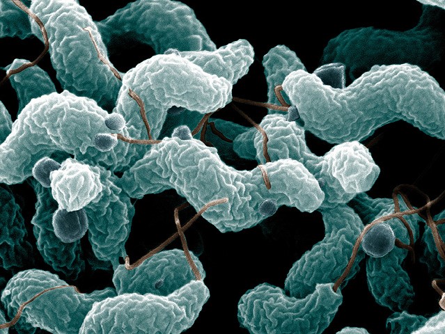
Campylobacter causes infections associated with faecal contamination of food or water.

They clinically presented with acute abdominal pain followed by watery diarrhoea with blood and leucocytes or dysentery.

Sometimes they may cause influenza-like prodrome of fever and generalized aching. Infections may present with rigors and sweating.

Nausea is common symptom. However, vomiting is not commonly seen.

Most of these infections are self-limiting. In severe disease antibiotic treatment is required.

Campylobacter species produce enterotoxins and cytotoxins.

These organisms are found in the intestine of poultry, pigs, sheep, goats, cattle and other animals.

The main cause of human infection are poultry, unpasteurized milk and contaminated water.

Faecal specimens are used for laboratory diagnosis of this infection. The specimen should be refrigerated at 2–8ºC if any delay to transport to the laboratory.

They can be stored up to 24 hours in refrigerator before processing as Campylobacter resistant to cold temperatures.

Microscopy

Campylobacters are small, spirally curved organisms with single flagellum at one or both poles.

They are motile bacteria. They show rapid darting motility.

Campylobacters are Gram-negative rods. When observed 1% basic fuchsin stained faecal smears, the organisms appear as wings of gulls and some appears in “S” shape or comma shape.

Faecal smears stained with basic fuchsin supply information for the presumptive diagnosis of Campylobacter enteritis.

Moreover, they are looks like spirochaetes as they become longer in the development process.

Filtration techniques are used to isolate Campylobacter species from faecal specimens.

Culture

Oxygen is essential for the growth of Campylobacters.

For the growth microaerophilic atmosphere containing 5% oxygen and 10% carbon dioxide is necessary. They grow well at 37–42°C.

Blood agar

Campylobacter jejuni and Campylobacter coli form non-haemolytic colonies on blood agar. They are droplet like colonies.

Improved Preston blood-free medium

coli produce small, creamy-grey colonies. They are moist and slightly raised. Swarming growth also can be occurred.

C. jejuni forms grey, moist colonies. They are flat and spreading colonies.

Biochemical Identification

Oxidase positive

Catalase positive

Latex agglutination tests are available for the diagnosis of Campylobacter.

PCR tests are also available for the differentiation and diagnosis of Campylobacter.

For epidemiological purposes DNA fingerprinting methods such as multi-locus sequence typing is used.

Complement fixation test and enzyme-linked immunosorbent assay (ELISA) are use to detect the infections by Campylobacter.

Treatment

Usually diseases caused by Campylobacter are self-limiting.

Sometimes very rarely patients require fluid and electrolyte replacement therapy.

Only in sever disease antibiotic treatments should be done. Erythromycin, Ciprofloxacin and fluoroquinolones are used for the treatment of the diseases caused by Campylobacter.

Prevention and Control

There is no vaccine available for the prevention. Proper waste disposal and personal hygiene is important.

Measures should be taken to reduce the number of Campylobacters and transmission of them in poultry industry.

Purification of water and heat treatment of milk can be done to prevent and control the Campylobacter infections.

Elimination of Campylobacters in carcasses can be done by γ-irradiation or chemical washes.

No responses yet

Leave a Reply