Epithelial tissues are one of major tissue type composed of continuous sheets of cells (one or more layers thick) that cover the body surface, line body cavities and hollow organs and also form glands.

There are two major types of epithelia.

  1. Covering epithelia

Cover all the outside surfaces of the body and line all the internal body cavities and tubes.

E.g. – Skin, Mucous membranes, Mesothelium

  1. Glandular epithelia

Secrete hormones and other products and found in glands.

The structure of epithelia is closely related to its functions, which include:

  • – Secretion
  • – Absorption
  • – Protection of underlying structures from dehydration, radiation, toxins and physical trauma.
  • – Regulation and excretion of chemicals between underlying tissues and the body cavity.

Characteristics of Epithelial Tissues

The cells of epithelium are densely packed together with minimum amount of intercellular matrix.

The upper surface of the epithelium is oriented towards the outside of the body or internal body cavity. The Basal surface usually lie on a basement membrane partly derived from underlying connective tissue.

Epithelium layers do not contain blood vessels. But innervated. Nutrients and waste are exchanged through neighboring connective tissues by diffusion.

Classification

Epithelial tissues can be classified according to morphological characteristics, which include;

  • Number of cell layers.
  • Shape of the component cells
  • Presence of surface specialization such as cilia, microvilli and keratin

According to the number of cell layers, there are three types of epithelia;

  1. Simple epithelia
  2. Stratified epithelia
  3. Pseudostratified epithelium

Simple epithelia consist of a single layer of identical cells resting on a basement membrane.

They are usually seen on absorptive or secretory surfaces and can be divided into three main types: simple squamous, Cuboidal and Columnar epithelia.

These types are named base on the shape of the cells, which differ from their functions.

Stratified epithelia consist of multiple layers of cells of various shapes. The cells of basal layers continuously divide and move cells towards the apical layer.

Stratified epithelia are mainly involved in protection of underlying structures by withstanding abrasions.

This can be divided in to four types: stratified squamous, cuboidal, columnar epithelia and transitional epithelium

Pseudostratified epithelium is a variant of simple columnar.

It is composed of one layer of columnar cells with nuclei at different levels. All cells reach the basement membrane but not the apical layer.

It usually contain goblet cells and cilia.

Based on the shape of the component cells, there are three types of epithelia.

  1. Squamous epithelia
  2. Cuboidal epithelia
  3. Columnar epithelia

If the epithelia is stratified, the shape of the outer most layer decide the classification.

Squamous epithelia are consist of flat, irregular shaped cells with centrally located nuclei. Their width is much greater than height.

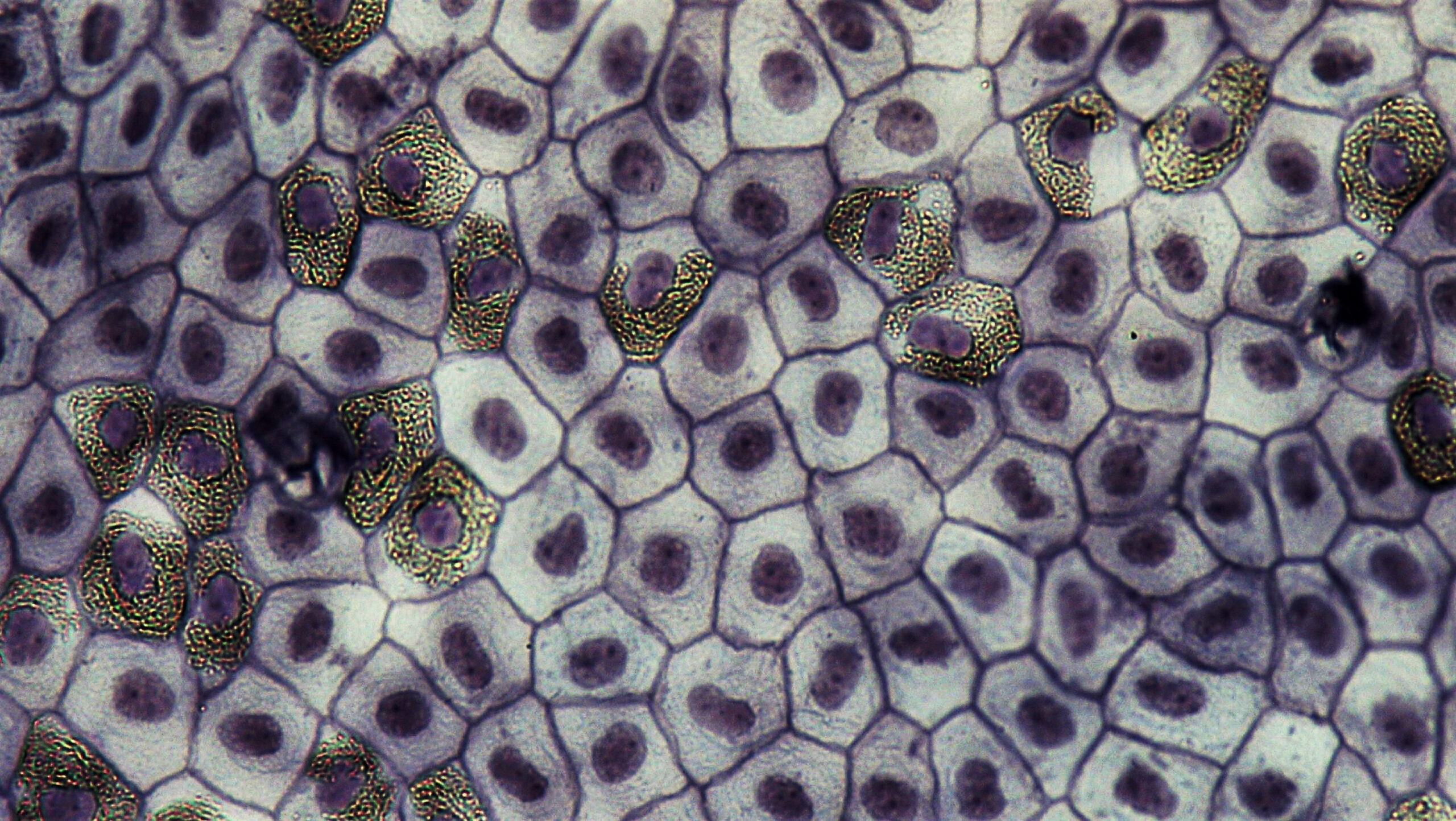
Cuboidal epithelia are consist of cube shaped cells. Cells have polygonal shaped surface and central round nucleus.

Columnar epithelia are consist of tall cells, appear as columns in sections. The nuclei are elongated and located towards the base in the center or to the apex. This epithelia may have cilia and microvilli.


Epithelial tissues can be classified according to the presence of surface specializations which are, microvilli, cilia & stereocilia and Keratin.

Microvilli are minute finger like projections, present on luminal plasma membrane. They are seen in cells specialized for absorption and increase the surface area. e.g.- Small intestine, proximal renal tubules.

Cilia are long, actively motile structures projects from the apical surface. They are easily seen by light microscope. Lining epithelium of the upper respiratory tract and fallopian tube contain cilia. Rhythmic movement of cilia moves mucous upwards in the respiratory tract. In fallopian tube, cilia propels the ovum from the ovary to the uterus.

Stereocilia are extremely long microvilli. They are readily visible by light microscope. Epithelium of epididymis and vas deferens contain stereocilia. Probably water absorption occurs by this specialization.

Keratin is found in areas susceptible to abrasion and water loss. Based on the presence of keratin which a intermediate filament protein, stratified squamous epithelium can be divided in to keratinized squamous and non- keratinized squamous epithelium.

Based on the number of cell layers and the shapes of component cells, there are eight types of epithelia;

  1. Simple squamous epithelium
  2. Simple cuboidal epithelium
  3. Simple columnar epithelium
  4. Pseudostratified columnar epithelium
  5. Stratified squamous epithelium
  6. Stratified cuboidal epithelium
  7. Stratified columnar epithelium
  8. Transitional epithelium

Simple Squamous Epithelium

This is composed of single layer of flattened cells resting on a basement membrane with centrally located, flattened nuclei. The cells fit closely together, forming a thin and very smooth membrane which facilitates for diffusion.

Common locations:

  • Endothelium of blood vessels and lymphatics (facilitate exchange of material between blood and tissue fluid.)
  • Mesothelium; serous membrane which line closed body cavities such as pericardium, pleura & peritoneum. (allow passage of tissue fluid in to and out of body cavities.)
  • Lung alveoli ( exchange of gases)
  • Bowman’s capsule (filtration in Bowman’s capsule)

Simple Cuboidal Epithelium

This consists of single layer of cube-shaped cells with central, round nucleus. The cells fit closely together lying on a basement membrane. Secretion, absorption and/or excretion are the functions of cuboidal epithelium

Common locations:

  • Glandular epithelium , e.g.-thyroid
  • Small excretory ducts of salivary glands and pancreas
  • Convoluted tubules of the kidney
  • Surface of ovary (germinal epithelium)

Simple Columnar Epithelium

This is formed by single layer of elongated cells on a basement membrane with basally located, elongated nuclei at the same level.

Some of simple columnar epithelia have surface specializations that suitable for a specific function.

Microvilli that cover the free surface of the columnar epithelium lining the small intestine provide a very large surface area for absorption of nutrients from the small intestine. This tissue type mainly involve in absorption and secretion.

Common locations:

  • Lining of entire digestive tract ;Small intestine, Large intestine, Gallbladder, Stomach

Goblet cells are modified columnar epithelial cells which synthesize and secrete mucin.

They are scattered among the cells of many epithelial linings of respiratory tract and digestive tract.

The mucin vacuole is bulged and the nucleus is compressed in a goblet cell.

Pseudostratified Columnar Epithelium

Pseudostratified columnar epithelium is composed of a single layer of columnar cells with nuclei at different levels. The cells differ in height and not all cells reach the apical surface.

The term pseudostratified is derived from the appearance of being multilayered. It usually contain and goblet cells and cilia.

This involves in Secretion and absorption as well as ciliated tissue moves mucus.

Common location:

  • Upper respiratory tract including trachea

Stratified Squamous Epithelium

This is composed of several layers of cells resting on a basement membrane. The basal layer vary from cuboidal to columnar.

Basal cells become flattened and then shed, when they grow towards the surface by dividing continuously. Nuclei become progressively condensed (pyknotic) also flattened before ultimately disintegrating.

This tissue type facilitates to withstand abrasions and occur a barrier function.

There are two types of stratified squamous epithelium based on accumulation of keratin:

  1. Keratinized squamous epithelium.
  2. Non-keratinized squamous epithelium.

Keratinized Squamous Epithelium

The outermost layer consist of tightly packed dead cells that have lost their nuclei and filled with a protein, keratin.

This layer act as a tough, water proof protective layer that prevents drying of the live cells underneath.

Common location:

  • Epidermis of skin

Non Keratinized Squamous Epithelium

Moist superficial cells are living. This protects moist surfaces which subjected to wear and tear, and prevents them from drying out. Protection and secretion are the main functions of this epithelium.

Common locations:

  • Mouth
  • Epiglottis
  • Esophagus
  • Vocal folds
  • Vagina
  • Ectocervix

Stratified Cuboidal Epithelium

This usually consist of only 2-3 layers of cuboidal or low columnar cells.

This tissue type provide a lining and is not involved in significant absorptive or secretary activity.


Common locations:

  • Ducts of sweat glands
  • Large ducts of exocrine gland
  • Parotid gland
  • Sub mandibular
  • Anorectal junction

Stratified Columnar Epithelium

This is composed of single layer of columnar cells on several layers of cuboidal cells. Secretion, absorption and protection are the main functions of this epithelium.

Common location:

  • Conjunctiva of eye
  • Some large excretory ducts (Salivary gland)

Transitional Epithelium (Urothelium)

This is composed of several layers of pear-shaped cells and features intermediate between stratified cuboidal and stratified columnar epithelium.

This epithelium found only in the urinary tract in mammals.

This epithelium is highly specialized to accommodate great degree of stretch and to withstand toxicity of urine.

When the urinary tract changes from non-stretched state to stretched state, the number of cell layers change from 4-5 cell layers to 2-3 cell layers.

Categories:

No responses yet

Leave a Reply