Introduction

Grocott’s Methenamine Silver Stain is a special staining method used in Histological diagnostic procedures. Grocott’s Methenamine Silver Stain is useful in the identification and screening of Fungi.

Moreover, this stain is useful in staining carbohydrates.

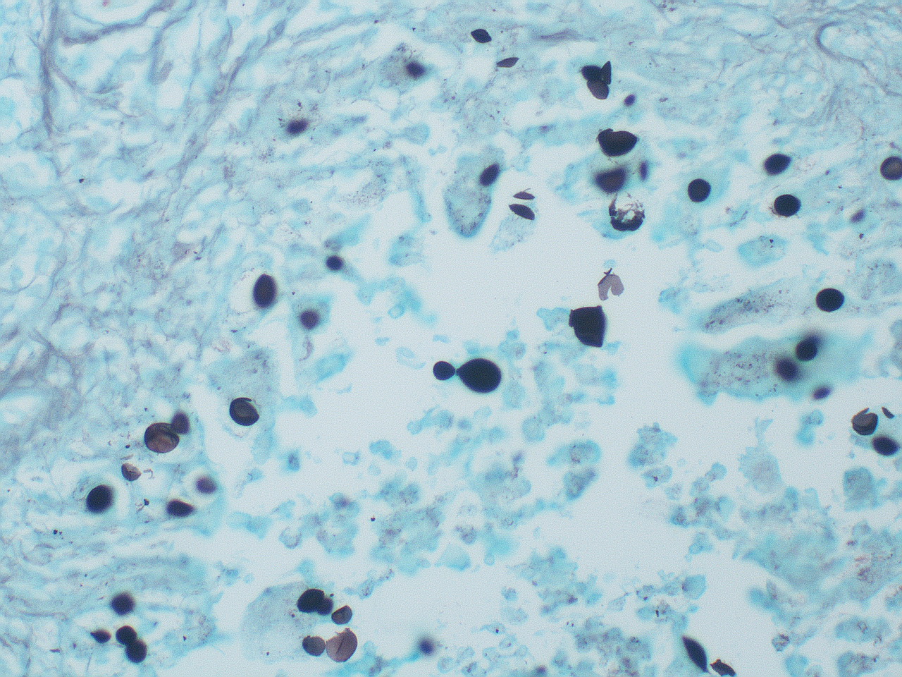
By using this stain yeast-like fungus can be identified.

This stain, stains all pathogenic and non-pathogenic fungi.

Mainly this staining is done for the identification of fungus such as Pneumocystis pneumoniae, Pneumocystis jiroveci, Aspergillus species, Candida species.

Here staining of the fungal wall is done by this stain.

Principle

Fungal cell wall is made up of polysaccharides.

These polysaccharides can be converted into dialdehydes, by oxidation of them.

These dialdehydes are detected by the silver solution.

The fungal cell walls are stained in dark brown or black in colour after staining by GMD stain and counter stain give green colour to the background.

For this staining procedure 5% aqueous chromium trioxide (chromic acid) solution, 1% aqueous sodium metabisulphite solution, 5% sodium thiosulphate (Hypo) solution is needed.

In addition, 1% Light green is used as counter stain.

Other than that, 0.2% aqueous gold chloride solution and 5% sodium tetraborate is also essential for the staining procedure.

Preparation of working solution

Solution A and solution B should be prepared separately.

For the preparation of solution A, add 18-20 drops of 10% Silver nitrate to 25ml of 3% hexamine.

Then these solutions should be mixed well.

For the preparation of solution B, mix 3ml of 5% sodium tetraborate with 22ml of Distilled water.

Then mix solution A and solution B well. Final volume is 50ml.

Method

  • First hydrate the tissue section by bringing them down to the water.
  • Then treat with chromic acid solution for 1 hour.
  • Next wash the section well using water.
  • After that add Metabisulphite solution. It is done for the bleaching purpose.
  • Then wash in tap water. Pre heat the silver solution and place the sections in that solution.
  • Leave the sections in silver solution for about 15 minutes.
  • After 15 minutes check the sections, whether they have got black.
  • Leave sections until they get black. After that rinse in water.
  • Apply gold chloride on sections for few seconds. Then rinse in water.
  • Next fix the sections using 5% sodium thiosulphate solution for few seconds.
  • Wash the sections and counter stain using 1% Light green solution for 1-2 minutes.
  • Then wash the sections well. Finally dehydrate, clear and mount the sections with DPX.

Purpose

It is useful in the identification and screening of Fungi

Results

Fungi, Pneumocystis and melanin stains in black colour.

Mucin and Glycogen parts stain in dark grey colour. Background stains in green colour due to light green solution.

Other

The staining procedure can be differed according to the condition of the stain.

Time of staining should be adjusted accordingly.

It is necessary to use a positive control slide of Pneumocystis jiroveci or similar organism.

Always results should be interpreted if the positive control has stained correctly. Under staining of tissue sections may cause false negative results.

Therefore, it is necessary to consider about the staining procedure and change them accordingly under a supervision of Medical Laboratory Technologist.

No responses yet

Leave a Reply