Learn about bacteria

Staphylococcus aureus


Staphylococci are Gram-positive cocci occurring in clusters. They can be cultured on normal nutrient media both aerobically and anaerobically. In 1880, Dr Alexander Ogston, first showed that a number of human pyogenic (pus forming) diseases were associated with a cluster- forming micro-organism. He was introduced the name ‘staphylococcus’ ( In Greek  language: staphyle mean ,bunch of grapes; kokkos mean grain or berry).

Staphylococcus aureus

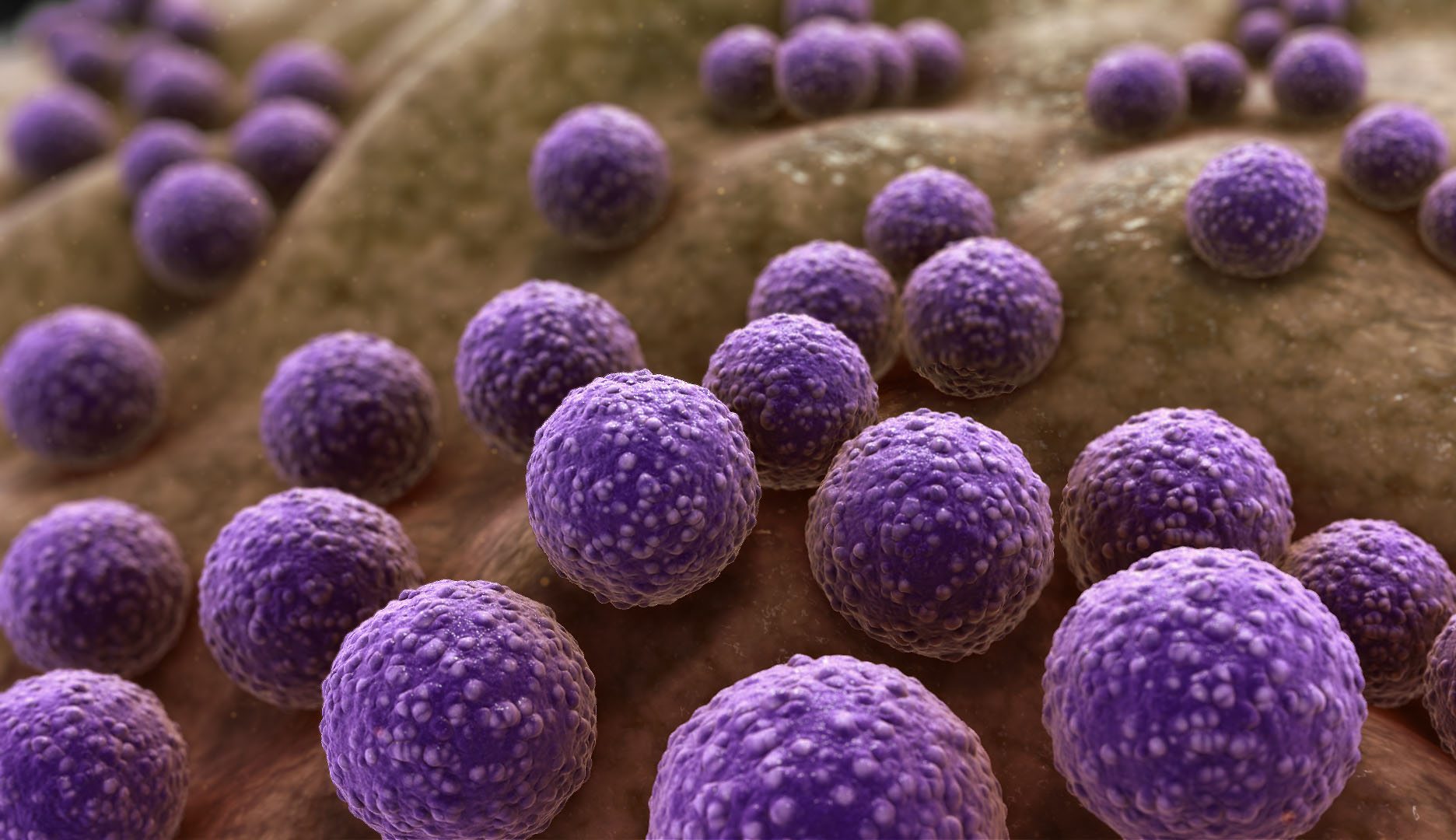
Now it uses as the genus name of facultatively anaerobic group. They may also can be found as part of the normal flora of other sites such as the upper respiratory tract, and are commonly present on animals. The major pathogen within the genus, S. aureus, causes a wide range of minor and major infections in human and animals. S. aureus causes suppuration, abscess formation, a variety of pyogenic infections and can even cause fatal septicemia. Pyogenic infections are Boils, Carbuncles, surgical site (wound) infection, abscesses (spinal), impetigo mastitis, blood stream infections, osteomyelitis, pneumonia (ventilator-associated) and Endocarditis. Toxin-mediated infections are Pemphigus neonatorum, toxic shock syndrome, Scalded skin syndrome and food poisoning.

A number of virulence factors are responsible for the clinical symptoms of infections by this pathogen. Extracellular enzymes and toxins contribute to its invasiveness and pathogenicity.

  • Coagulase: Clots plasma, interferes with phagocytosis, facilitates spread in the tissues.
  • Haemolysins: Lyze red cells.
  • Leukocidin: Kills leucocytes.
  • Fibrinolysin: Digests fibrin.
  • Lipase: Breaks down fat.
  • Hyaluronidase: Facilitates spread in tissues by destroying hyaluronic acid (component of connective tissue).
  • Protein A: Antiphagocytic (prevents complement activation).
  • Enterotoxins (heat stable): Cause food-poisoning (particularly vomiting).
  • Toxic shock syndrome toxin-1: Shock, rash, desquamation of skin.
  • Epidermolytic toxins A and B: Generalized peeling of the skin.
  • Chemotaxis inhibitory protein: Inhibits migration and activation of neutrophils.

laboratory specimens


Pus and swabs from infected sites, sputum, cerebrospinal fluid, and blood for culture are useful for the diagnosis of this organisms. Feces, vomit and the remains of food when food poisoning is suspected are also used as laboratory specimens.

Growth & Colony Morphology

Staphylococci grow well aerobically and in a carbon dioxide enriched atmosphere. Most strains also grow anaerobically, but less well. Temperature range for growth is 10–42ºC, with an optimum of 35–37ºC.

Staphylococcus_aureus_colony morphology


S. aureus produces yellow or cream or occasionally white 1–2 mm in diameter colonies after overnight incubation on blood agar. Some strains are beta hemolytic when grown aerobically on blood agar and chocolate (heated blood) agar. Colonies are slightly raised and easily emulsified. 


Staphylococcus_aureus
Staphylococcus on MacConkey agar


On MacConkey agar S. aureus produce Smaller 0.1–0.5 mm colonies after overnight incubation at 35–37ºC. Most strains are lactose fermenting.


Mannitol salt agar is a useful selective medium for recovering S. aureus from fecal specimens when investigating staphylococcal food-poisoning. It can also be used to screen for nasal carriers. Staphylococcus aureus ferments mannitol and is able to grow on agar containing  sodium chloride. Mannitol salt agar containing  sodium chloride (plus 4 mg/l methicillin) is recommended, particularly for isolating MRSA strains.

Staph aureus on MSA

The organisms are non-spore forming, non-motile and usually non-capsulate.  The main distinctive diagnostic features of S. aureus are,

  • Production of an extracellular enzyme, coagulase, which converts plasma fibrinogen to fibrin, aided by an activator present in plasma.
  • Production of thermo stable nucleases that break down DNA.
  • And also Production of a surface-associated protein known as clumping factor or bound coagulase that reacts with fibrinogen.

Bio chemical tests for the diagnosis of Staphylococcus Aureus

1. Catalase tests

Catalase Test


This test differentiates Staphylococci which are catalase positive from Streptococci which are catalase negative.

Note: Slide coagulase test is not recommended because of the risk of contamination from active bubbling.

2. Coagulase test

Coagulase Test


These isolates have either thickened cell walls (important to reduced susceptibility) or the vanA gene (important to fully resistant), and can be difficult to detect in the routine diagnostic laboratory.

Other important Features


There is no vaccine against staphylococci. Cleanliness, frequent hand washing, and aseptic management of lesions, proper sterilization of instruments, good aseptic techniques when handling surgical instruments help to control spread of S. aureus.


There are commercially available agglutination tests to identify S. aureus. Several latex agglutination test kits have been developed to identify S. aureus based on the detection of clumping factor, and, or protein A.

latex agglutination test

Other pathogenic Staphylococcus species

They also called as coagulase negative staphylococcus (CNS). Staphylococcus saprophyticus (S. saprophyticus) causes urinary tract infections in sexually active female. Staphylococcus epidermidis cause  usually  endocarditis and bacteremia following infection of cannulae, indwelling catheters, shunts or other appliances positioned in the body. Infections are difficult to treat due to the resistance of S. epidermidis to many antimicrobials.

Microscopically, S. saprophyticus and S. epidermidis resemble S. aureus.

No responses yet

Leave a Reply