Introduction

Congo Red stain is useful for the demonstration of Amyloids in tissue sections.

Amyloids are the abnormally accumulated proteins in tissues. They are abnormal insoluble filaments.

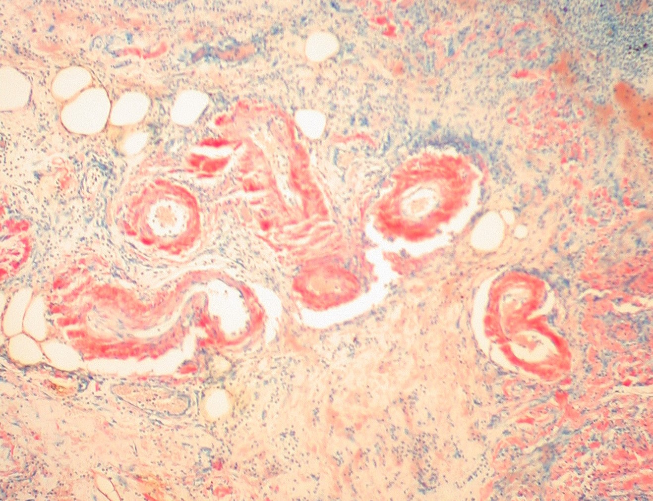
Amyloids deposited in the tissues damage the structure and function of the tissues and cause fatal conditions when they deposited in important major organs.

There are different types of Amyloids such as primary amyloids, secondary amyloids, tumor-associated amyloids and myeloma-associated amyloids.

The gold standard technique for the diagnosis of amyloids deposited in tissues is Congo Red method.

Electron microscopy also used for this diagnosis procedures.

These sections also can be viewed under light microscope.

In hematoxylin and eosin (H&E) stained tissue sections amyloid can be seen as extracellular, eosinophilic, amorphous substances.

However, it is not easy to differentiate them by staining with hematoxylin and eosin stain.

Therefore, Congo red stain is useful for that.

The molecular formula for Congo red is, C32H22N6Na2O6S2.

It is a sulfonated azo dye which is symmetrical. It contains hydrophobic center.

Furthermore, it is a linear molecule due to the two phenyl groups which have bound by a diphenyl bond.

Congo red is also considered as a fluorescent dye.

For the staining of amyloid by Congo red, linearity of the dye molecule and β-pleated sheet configuration is important.

Principle

Congo Red is an acidic stain. It composed of two identical halves.

In this molecule each half has a phenyl ring which has bound to naphthalene moiety by a diazo group.

These two phenyl groups bound by a diphenyl bond.

Because of that this dye molecule has a linear structure.

Amyloids are stain by Congo red by making hydrogen bonds between dye molecules and amyloids.

Other components in the tissue are stained due to the electrochemical bonds.

  • For the staining procedure Congo red solution, Carazzi’s Haematoxylin, 1% Potassium Iodide and 70% alcohol is necessary.
  • For the preparation of Congo red solution 3 ml of 1% Congo red in 4% Potassium citrate solution should be mixed with 1 ml of Glycerin.
  • These two solutions should be mixed well together just before starting the staining.
  • That means the Congo red solution should be prepared freshly.
  • First bring the sections down to the water for hydration. Then stain with the freshly prepared Congo red solution for 10 minutes.
  • These slides should be incubated at 56°C incubator.
  • Then wash with water. After that cover the slides with 1% Potassium Iodide solution for 3-5 minutes.
  • 1% potassium Iodide is considered as a mordent.
  • Then wash the sections with water.
  • Next stain with Carazzi’s Haematoxylin for about 3 minutes.
  • Blue the sections in tap water for 10-20 minutes.
  • Then treat again with 1% Potassium Iodide solution for several minutes.
  • This step is optional.
  • Then differentiate the sections in 70% alcohol.
  • Finally dehydrate, clear and mount with DPX.

Purpose

This method is useful for the staining and demonstration of Amyloids in tissue sections.

Results

Amyloids stain in Brick Red colour.

Nuclei stain in blue. Background stain in pale pink colour or it can be clear.

Moreover, Elastic fibers and Eosinophil granules also stain in Red colour.

The staining procedure can be differed according to the condition of the stain.

Time of staining should be adjusted accordingly.

It is necessary to use a positive control slide.

This staining procedure can be used for formalin fixed, paraffin embedded tissues and Frozen sections as well.

Congo red has a carcinogenic property.

Therefore, it is necessary to take recommended actions when doing the staining procedure.

For this staining the thickness of the sections should be 5-6 µm.

No responses yet

Leave a Reply