Introduction

Giemsa stain is named for the honor of Gustav Giemsa. Giemsa stain is a widely used nucleic acid stain.

Mainly used for the cytogenetics and diagnosis of malaria and other parasites in histological sections.

Identification of protozoa is mainly done using H&E stains.

However, Giemsa stain is the best method for identification of protozoa.

Moreover, Giemsa stain is used for the staining of blood and bone marrow films.

Giemsa is a mixture of staining solutions namely Methylene blue, Eosin and Azure B.

There is a commercially available Giemsa powder for the preparation of stain.

Principle

Giemsa stain is composed of both basic dyes and acidic dyes.

Basic dyes are methylene blue and azure b. Acidic dye is eosin.

Basic components stain with acidic dyes and acidic components stain with basic dyes.

Basic dye binds to acidic components of the nucleus and stains the nucleus and other acidic compounds in blue colour.

Acidic dye eosin, bind with cytoplasm, cytoplasmic granules and other basic components and produce red or pink colour.

The pH value of the stain should be maintained to bind the dyes to the cell components.

According to the fixatives used, pH adjustment should be done.

Giemsa stain for parasites in histological sections

  • For the preparation of Giemsa stock solution, First dissolve 4 g of Giemsa powder in 250 ml of Glycerol at 60°C.
  • Then shake well. Add 250 ml of methanol.
  • Shake well. Then allow the mixture to stand for 7 days. Then filter before use.
  • For the preparation of working Giemsa solution, measure 4 ml of stock Giemsa solution and mix it well with 96 ml of Acetate buffered distilled water.
  • Final pH should be 6.8.
  • First bring deparaffinized sections down to the water through a graded alcohols series to water.
  • Rinse in pH 6.8 buffered distilled water for few minutes.
  • Stain in filtered working Giemsa solution for overnight or stain the sections for 20 minutes at 56°C until the section gets dark blue.
  • Rinse the sections in distilled water.
  • Differentiate in a very weak Acetic acid solution (0.5% aqueous acetic acid solution) until sections get pink in colour.
  • Rinse in distilled water. Dehydrate, clear and mount with DPX.

Results

Protozoa and some other microorganisms stain in dark blue.

Background stains in pink colour.

Nuclei stains in blue colour. Azurophilic granules stain in blue colour.

May–Grunwald–Giemsa Stain for Blood and Bone marrow staining

  • First dry the blood and bone marrow films in the air.
  • Then immerse the films in a jar containing methanol for 5–10 minutes.
  • For bone marrow films, allow the bone marrow films for a longer time for better drying.
  • Then immerse in a jar containing methanol for 15–20 minutes for fixation.
  • Blood and bone marrow films should be fixed as soon as possible after they have dried.
  • Methanol is the choice of fixative used for blood and bone marrow staining with Giemsa stain.
  • Then transfer the fixed films to a Giemsa stain containing jar.
  • Here working solution should be prepared freshly before use by adding same volumes of stock Giemsa solution and buffered distilled water.
  • Allow the film to stain for 15 minutes. Then transfer the films into a staining jar containing Giemsa stain without washing with water.
  • Keep the slides in staining jar about 10-15 minutes.
  • This working staining solution should be prepared by adding one volume of stock Giemsa to nine volumes of buffered water at pH 6.8.
  • After that transfer the slides to a jar containing buffered water at pH 6.8.
  • Then wash the slides in buffered water rapidly.
  • It is necessary to change the buffered water three to four times.
  • Next keep the slides undisturbed in water for about 2-5 minutes.
  • It is for the differentiation. This step should be done under the inspection.
  • The wet slides should be inspected under the low power of the microscope to check the differentiation and better staining.
  • When this differentiation step is completed, keep the slides upright to dry.

Fixative is not critical in histopathological diagnosis.

However, B5 or Zenker’s solution is more preferred.

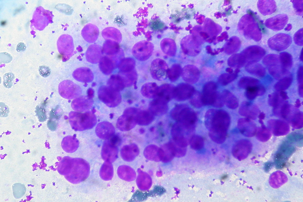
3 µm paraffin sections are preferred for Giemsa straining.

Working Giemsa stain should be prepared shortly before use.

Blood and bone marrow films should be fixed as soon as possible after they have dried.

If they are left unfixed at room temperature for more time, background drying artefacts can be seen after staining.

Avoid the contact of blood and bone marrow films with water before fixation is completed.

Instead of methanol, ethanol also can be used. Absolute ethanol should be used for this purpose.

However, methanol is the choice of fixative. Methanol should be completely free of water.

Because little amount of water can affect the appearance of films and higher amount of water contamination may lead to gross changes.

Finally, it may cause wrong diagnosis. When preparing smears, it is necessary to use detergent free slides.

If not the cell morphology may affected.

No responses yet

Leave a Reply