The word ‘sidero’ means iron, and ‘blasts’ mean immaturity and the name ‘sideroblastic anemia’ implies the presence of iron, in dysfunctional or immature red cells, leading to anemia. 

Etiology

Sideroblastic anemia occurs due to a defect in protoporphyrin synthesis, which is an essential step in haem production.

The causes can be congenital or acquired . 

Congenital sideroblastic anemia is caused by a genetic mutation which follows an X-linked pattern of inheritance.

This makes boys more prone to get X-linked sideroblastic anemia.

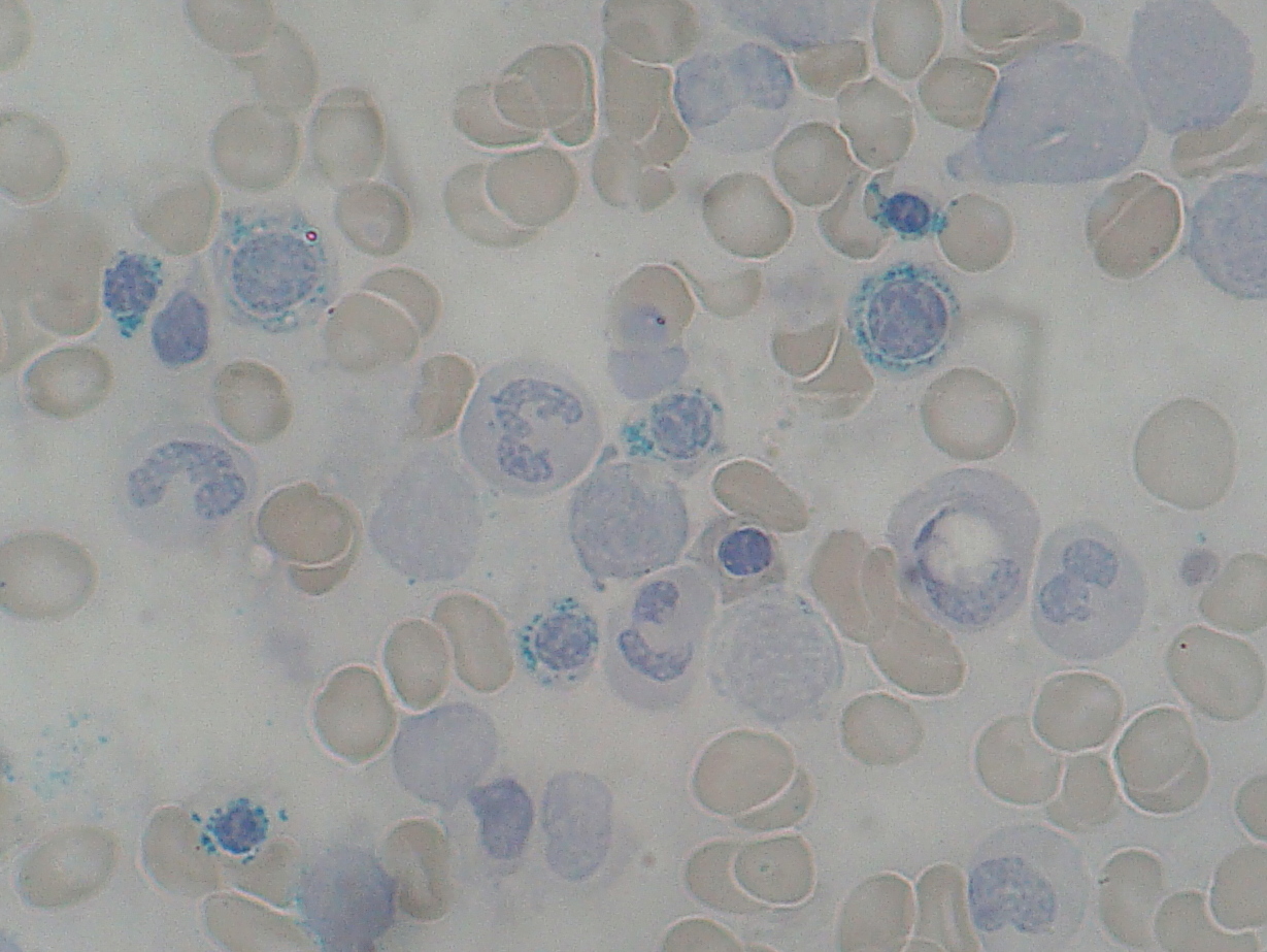
This particular genetic mutation causes Iron build-up in the red cells. 

Acquired causes mainly include excessive alcohol usage, nutritional deficiencies like vitamin B6 and folate, which affect the mitochondria’s ability to produce haem.

Other causes of the acquired form of disease include lead poisoning, treatment of TB with isoniazid, zinc overdose, and myelodysplastic syndrome. 

Pathophysiology

All the above mentioned situations render the bone marrow unable to use Iron even if Iron is readily available.

This causes deposition of excessive iron in the immature red cells, while causing an apparent iron deficiency in the bone marrow.

Signs and Symptoms

Pallor and tiredness, the major features of anemia will be there along with diarrhoea.

Due to the iron buildup in sideroblastic anemia, there can be organ damage too. The liver, heart, kidneys, spleen are the most susceptible organs.

The usual investigations done are full blood count, blood picture, and iron studies. 

While interpreting full blood count results is that while the mean corpuscular volume will be normal or lower in congenital causes, acquired causes will lead the MCV to be normal or higher. 

There can be some special features evident in the blood picture.

The most important feature is what gave this disease its name; the ring sideroblasts.

Immature red cells will contain iron deposited in a ring around their nuclei.

This can be seen in bone marrow histology too. 

Some of the other features in blood picture are Pappenheimer bodies, which are the iron depositions inside the mitochondria, and basophilic stippling, which is caused by the degraded ribosomes which indicates ineffective erythropoiesis. 

While the serum iron and ferritin will be high, the total iron binding capacity will be low. 

Treatment

Provide the deficient nutrients (vitamin B6, folate) if there is a nutritional deficiency.

To remove the excess iron iron chelating agents can be given. In severe cases blood transfusions or liver or bone marrow transplants have to be done.

Watch Video ...

Categories:

No responses yet

Leave a Reply